ankle fractures and Lisfranc injury clinical cases
June 14, 2020
by orthohub
These cases are are to get you to start thinking and questioning your beliefs ahead of the next webinar on ankle fractures and Lisfranc injuries.
We are joined for this webinar by:
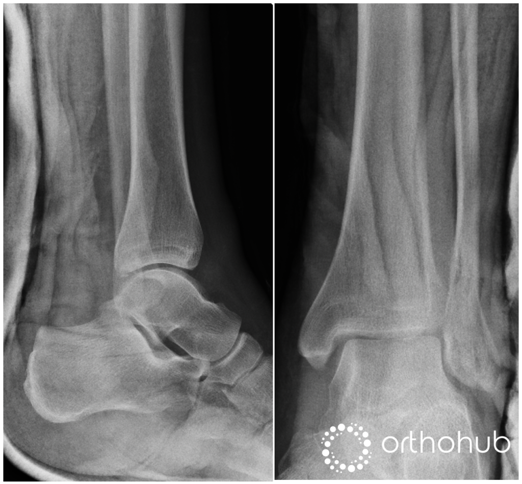
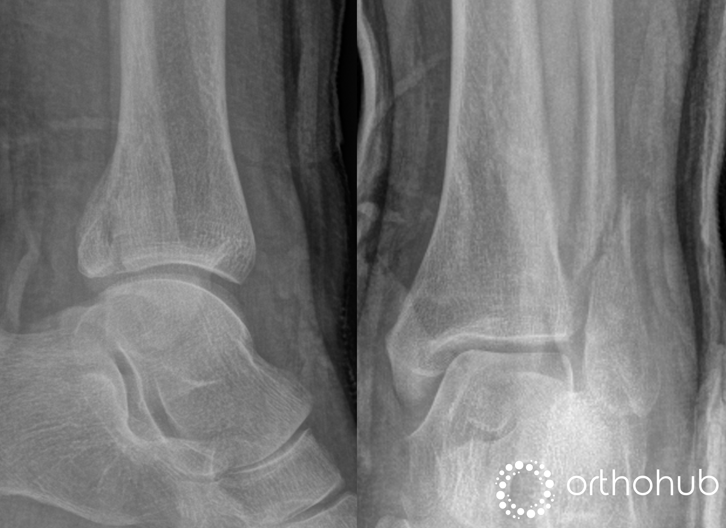
43 yrs male publican tripped over a step. Inversion injury to left ankle. Seen in ED and put in a backslab (injury view not available). The ED records report that he was diffusely swollen and painful medially.
No fractures are seen around the ankle on XR
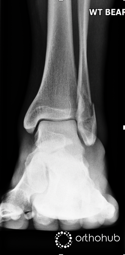
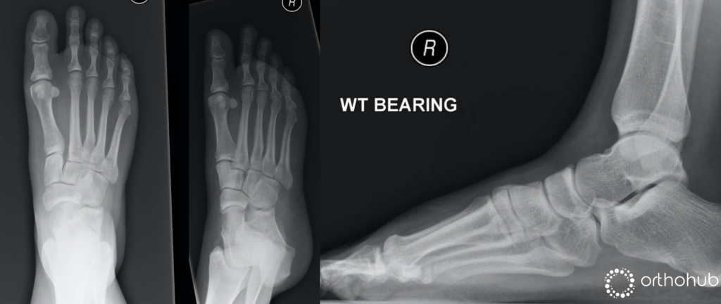
The treating doctor decided to do a weight-bearing view in cast:
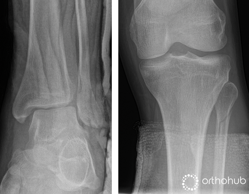

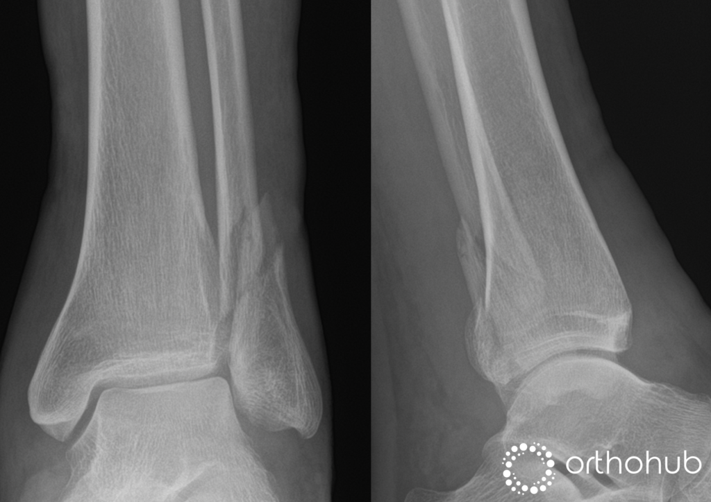
32 yrs male, sustained a twisting injury to his left ankle 2-days ago and sustained the injury above.
He was put in a backslab in ED and is now in fracture clinic.
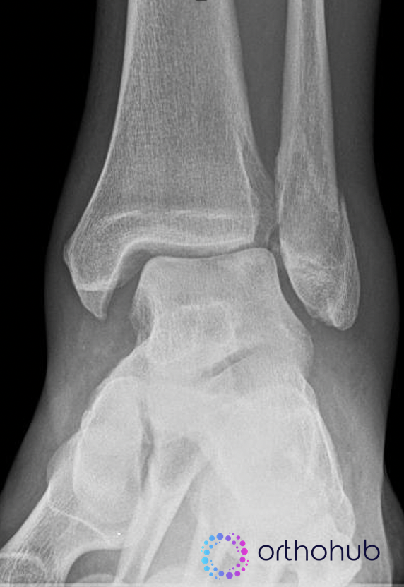
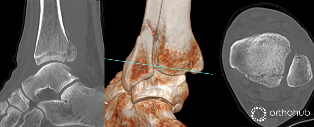
A 24 yrs car salesman sustained this injury while playing football. The doctor in fracture clinic removed the backslab and took this weight-bearing X-ray (Image 1). The patient is happy to go with whatever treatment you recommend.
While you were pondering what to do, the helpful SHO goes and gets a CT of the ankle anyway (Image 2).
This case ends up on your trauma list for fixation. Regardless of your initial feelings on the case (discussed above), you've agreed to carry out ORIF (imagine one of the other consultants you owe a favour to has asked you specifically).
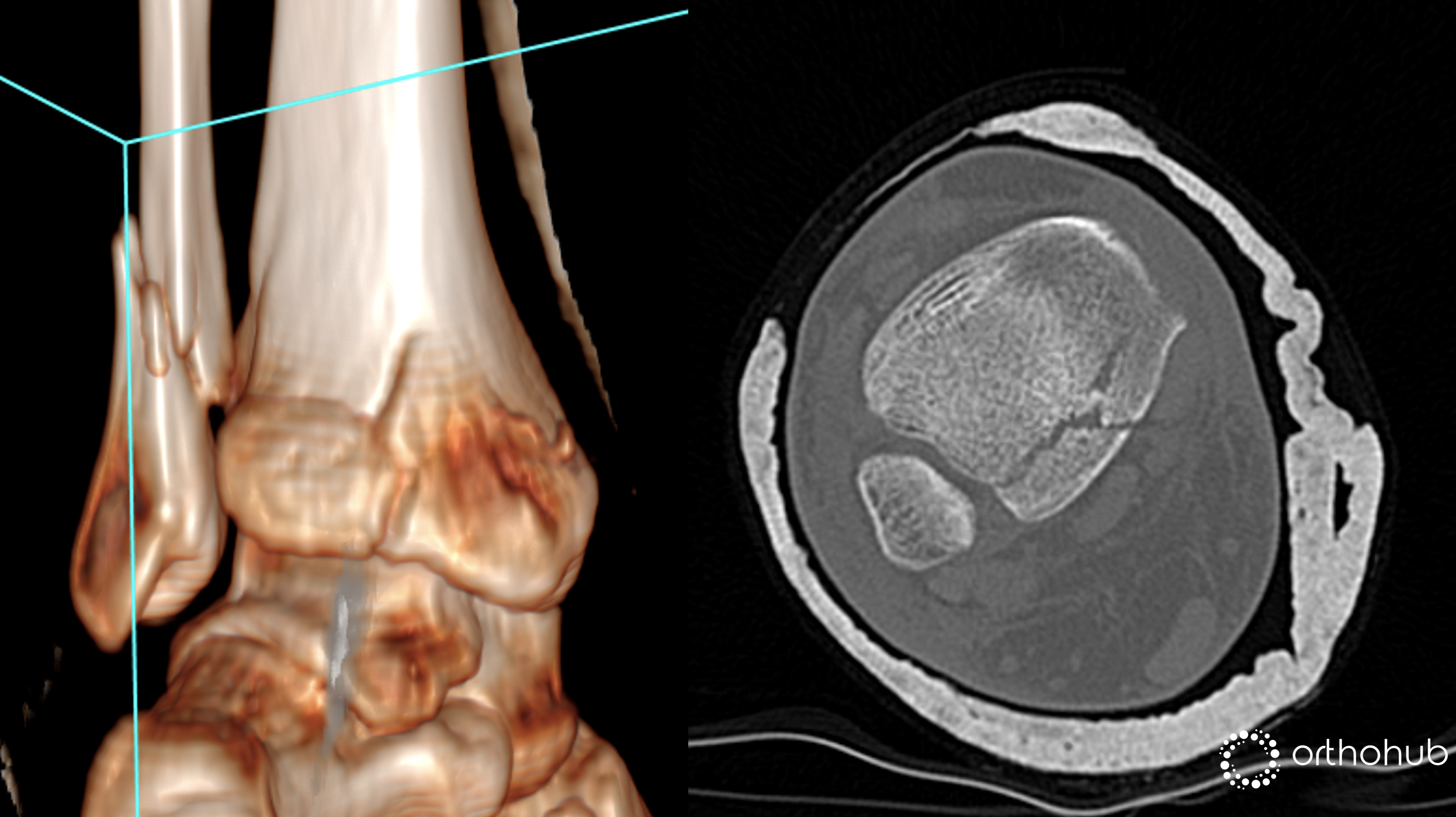
54 yrs female solicitor twisted her ankle getting out of the car. Plain X-rays and CT are shown.
Lyndon Mason explains how to use the CT scan to plan the surgical treatment of the posterior malleolus. Based on this you can select your surgical approach. Lyndon then talks us through how to perform the posterolateral, medial posteromedial, and posteromedial approach to the posterior malleolus.
29 yrs, fit and healthy store manager presents to fracture clinic having tripped whilst walking a week ago. He's complaining about right midfoot pain.
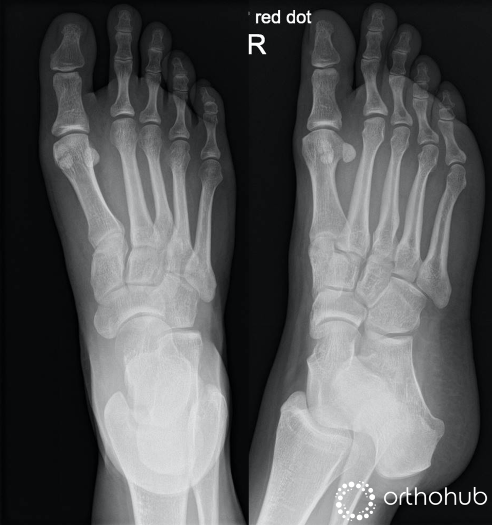
She is able to weight bear in a boot but she finds it painful. Clinically she is painful and swollen over the midfoot. Some midfoot swelling but no plantar ecchymosis.

Please do post your thoughts about these cases in the comments section below.
For the answers to these questions and much more join our webinar this week, Wednesday at 8pm BST.

Good cases
I'm interested to see how aggressive the faculty are in weightbearing the examples you had with a lateral and posterior malleolar fractures in the context of a normal weightbearing x-ray. I'd assume you'd just get them going in a boot - but i often feel a little apprehensive about doing this and keep a closer eye on them!
Good staff!!I just replied with our practice in my DGH which based by our foot ankle team iy looks work ,thanks still not sure if suspected lisfranc should we get this package straightaway ie WB XRAY PLUS CT or that should be sequntial !!
Hi Waleed, join us live on the webinar tonight and we will go through the answers to those questions.
We will post a video response too over the next week.
Great set of cases; looking forward to the discussion tonight!
Thank you for such a great teaching
Good cases
Very helpful to look through these cases indeed. Looking forward to lis Frank injuries discussion. Many thanks
Kind regards
Olga Jones
These are very common cases and have seen varied treatments. I would like to attend webinar and listen to expert on current evidence.
Great hopefully you will register and join us live tonight. Otherwise watch it later here ankle fractures and lisfranc webinar
Also the panel will be recording videos to answer the questions raised in these cases.
I am looking forward to seeing these topics being discussed tonight myself, great cases!
Great cases, thanks. With lots of embedded messages!
Glad you enjoyed them. Mike B